
Looking with atomic precision may be crucial to a materials researcher, no matter whether she/he wants to study organic solar cells, cement, or optical circuits. Hence, microscopes using electrons instead of light are the tool of choice, for robust materials at least. Sensitive materials, however, are damaged by illumination with energy-rich electrons. At Karlsruhe Institute of Technology (KIT), an electron microscope has now started operation, which reaches a high resolution with “soft” electrons.
“The new multifunctional electron microscope complements existing instruments at KIT and opens up new research horizons,” says Professor Dagmar Gerthsen, Head of the Laboratory for Electron Microscopy (LEM) of KIT. “The new microscope combines various tools in one instrument and can be used to obtain complementary information about a sample in a few working steps.”
Usually, the resolution of electron microscopes is improved by increasing the energy of electrons. As a result, however, the electrons may modify or even destroy structures in the sample to be investigated. The new microscope uses electrons of comparably low energy of about 30 keV and can be operated as a scanning electron microscope [Textfeld: Inner structure of carbon nanotubes with catalyst particles (top) and surface topography (bottom). (Photos: Cheng Sun/KIT)] (SEM) or scanning transmission electron microscope (STEM). Hence, it is suited for studying both the structure inside a sample as well as its surface topography. Nominal resolution of the instrument is about 0.3 to 0.6 nm (STEM and SEM, respectively), which corresponds to 3 to 6 atomic radii. In this way, structural properties in materials can be characterized and correlated with important functional properties in order to improve or understand major functions, such as the efficiency of solar cells, chemical activity of catalysts, or potential toxicological impacts of nanoparticles in biological cells.
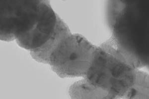
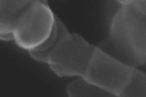
Inner structure of carbon nanotubes with catalyst particles (top) and surface topography (bottom). (Photos: Cheng Sun/KIT)
The microscope allows for the simultaneous investigation of samples with different detectors. “This yields new degrees of freedom in our investigation, which help us more than resolution alone,” explains Dr. Erich Müller of LEM at KIT. Various interactions of the electrons with the sample are used to provide experts with new findings relating to surface or volume properties of the sample. By X-ray analysis, also chemical composition of the samples can be determined. A special camera for imaging the diffracted transmitted or backscattered electrons allows conclusions to be drawn with respect to the crystalline structure of the material studied. “With a single instrument, we can now determine chemical and physical properties of samples much more comprehensively and are given insight in the atomic structure.” In the past years, the scientists of LEM configured the new microscope together with the manufacturer FEI. It is now the first instrument of this type delivered worldwide.
An integrated milling tool opens up additional research options for the nanoworld: A focused ion beam, FIB for short, can dig nanometer-sized trenches into the sample and, thus, uncover “hidden” layers below the sample surface. At interesting points, cross sections can be produced with high accuracy. The combination of high-resolution SEM or STEM with FIB and chemical analysis in one instrument makes the new microscope a key technology for fundamental and applied research to study the effects of nanoscale structures on material properties.
The Laboratory for Electron Microscopy of KIT executes own research projects, but also offers electron microscopy services to customers from industry and research. It operates a range of nine electron microscopes that offer optimum insight for a variety of problems. The flagship is an FEI Titan with a resolution of up to 0.07 nm. Research activities focus on the areas of solid-state physics, materials sciences, nanotechnology, chemistry, and biology. Another main activity is the development of methods for electron microscopy and electron optics.
More information:
https://www.lem.kit.edu/192.php (in German only)
Video on the LEM of KIT:
https://www.youtube.com/watch?v=7sFynh5TnAM (in German only)
In close partnership with society, KIT develops solutions for urgent challenges – from climate change, energy transition and sustainable use of natural resources to artificial intelligence, sovereignty and an aging population. As The University in the Helmholtz Association, KIT unites scientific excellence from insight to application-driven research under one roof – and is thus in a unique position to drive this transformation. As a University of Excellence, KIT offers its more than 10,000 employees and 22,800 students outstanding opportunities to shape a sustainable and resilient future. KIT – Science for Impact.