
From the Lecture Hall to the Operating Room
KIT Students Experience Medical Technology in Neurosurgery at Karlsruhe City Hospital
At 7:30 a.m. on a Tuesday in May, the workday for Professor Uwe Spetzger, Director of the Department of Neurosurgery at Karlsruhe City Hospital, has long since begun. He has already routinely completed his rounds and the preoperative briefing. But this is not a day like any other. For 15 students from Karlsruhe Institute of Technology (KIT), too, this day begins unusually – because today they will observe brain tumor surgery.
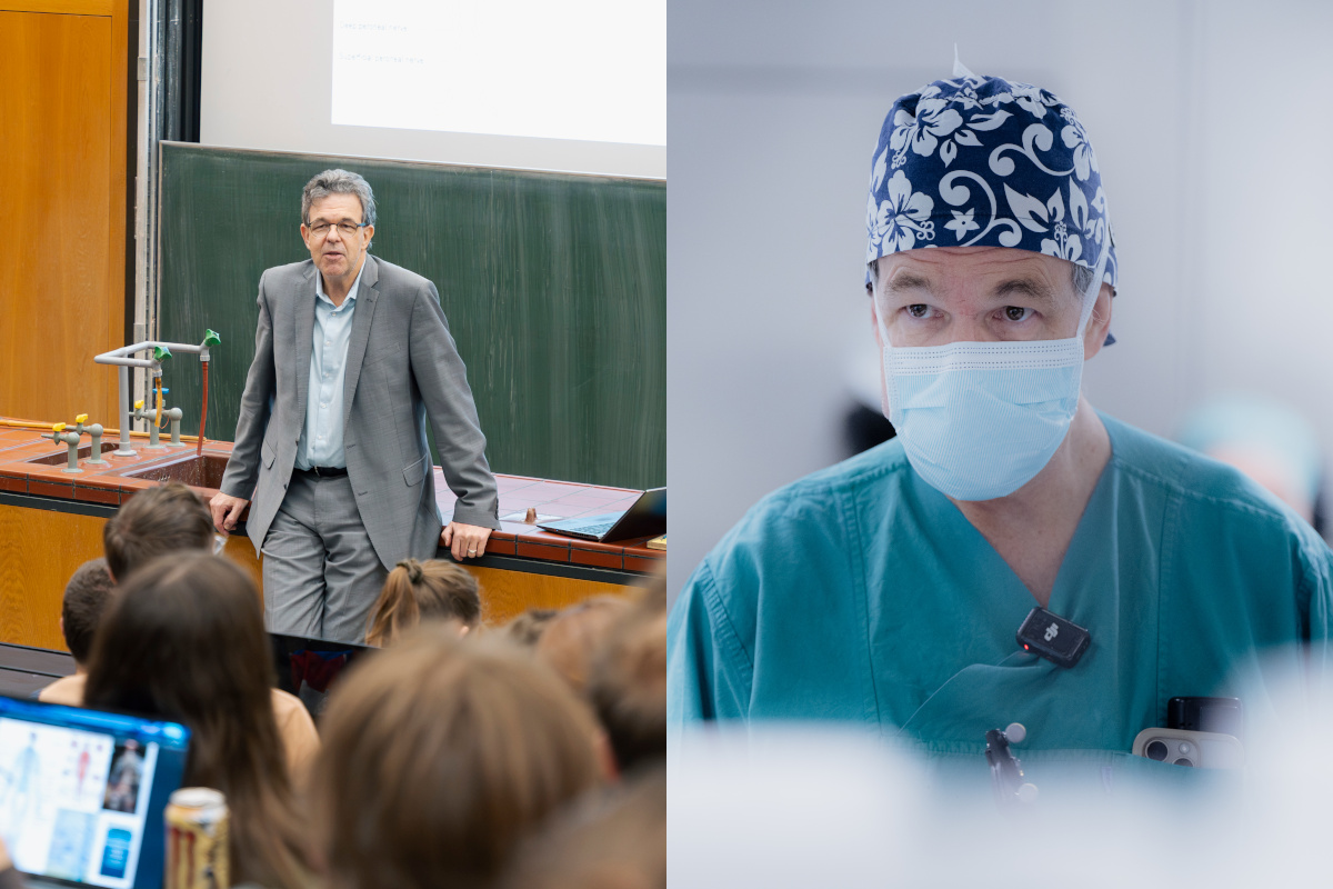
Spetzger works closely with KIT. For 18 years, the neurosurgeon has been an honorary professor at the Institute for Anthropomatics and Robotics at KIT and, every semester – usually after a morning surgery – he teaches the course “Introduction to the Brain and Central Nervous System.” “We usually start on time, but if an emergency comes up, that takes priority, of course,” says Spetzger. In the back row, a student mutters, “Lecturer and neurosurgeon – isn’t that pretty stressful?”
Medical technology, computer science, materials science, physics, mathematics – students from a wide variety of majors sit in the packed lecture hall with its ’70s charm. “You won’t graduate from here as medical students, but you should learn something about the anatomy of the brain and the nervous system,” Spetzger tells the audience. Master’s students in Biomedical Engineering have the opportunity to experience the technical systems they know from theory and the lab in practice – in Spetzger’s operating room.
“Neurosurgery is a high-tech discipline. I want to give students a real-world understanding of this technology. In our collaboration with major MedTech companies, we’ve seen that technologies are sometimes developed that are far away from clinical reality and do not work in practice. That’s why it’s important to involve students early on,” says Spetzger.
Preparation Is Everything
The group of students meeting in front of the Neurosurgery Department’s office in May has this opportunity. Sanne, a master’s student in electrical engineering specializing in biomedical engineering, already observed a surgery last winter semester: “Last time, a patient with a pituitary tumor was operated on through the nose. This time, we’ll see how the skull is opened – it’s going to be extremely exciting!”
Before heading into the operating room, Spetzger discusses the case with the group in the neurosurgery library. There’s just enough room for everyone to sit. All eyes are fixed on the large screen displaying numerous radiological images. Dr. Till Brombach, senior physician in neurosurgery at Karlsruhe Neurocenter, presents the case: The patient is 80 years old; the brain tumor is located in front of the motor cortex, is growing rapidly, and is very likely malignant. Without surgery, the woman would die within a few weeks. Upon hearing this diagnosis, the room falls silent for a moment.
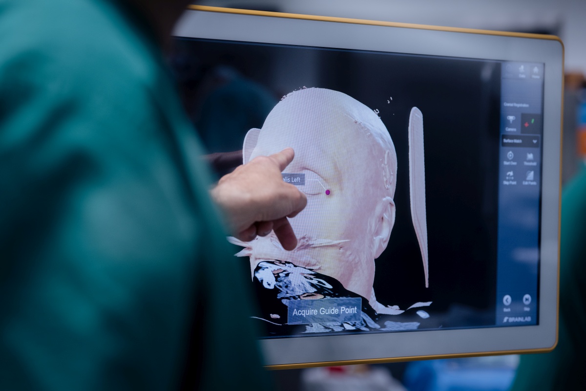
“To ensure we know exactly where we are during surgery, we work with MRI datasets that reveal the fine structures of the brain. In addition, we have CT scans that primarily show the bony landmarks. From both datasets, we generate a 3D model in which important areas are marked – a kind of navigation map that makes the procedure faster and more precise.” Uwe Spetzger leans against a cabinet in the background and adds: “Precise planning and intraoperative navigation are the be-all and end-all in neurosurgery. The navigation points used when surgically opening the skull must be as precise as possible so that as little healthy brain tissue as possible is cut through.”
Before heading into the OR, he gives a brief introduction: “You can’t go far wrong – just don’t touch anything and don’t contaminate anything. And if anyone isn’t feeling well, just sit down on the floor – that reduces the distance of a potential fall. While there’s no better place to faint and hit your head than in a neurosurgical operating room, it’s always about minimizing risk.” And with that, the group heads toward the operating room, first through the changing area.
Navigation Accurate to the Millimeter
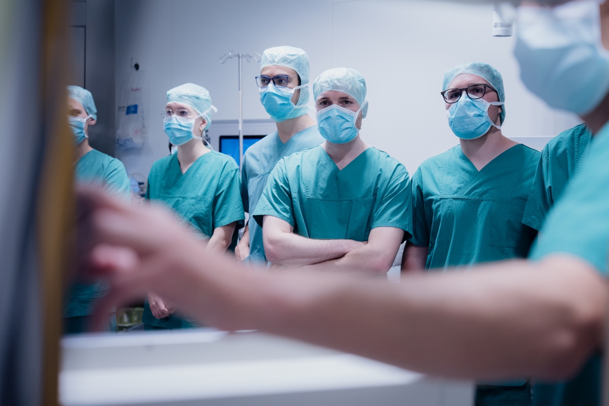
Wearing green surgical scrubs, surgical caps, masks, and with disinfected hands, everyone is allowed into the area that non-medical staff would normally never enter. The patient is already under anesthesia in the operating room, while every step of the preparation is carried out with practiced precision. Since space is limited, the students are divided into two groups – one group stands in the operating room, while the other observes the procedure through a large glass window.
Brombach explains how the head is secured in a head frame. Attached to it is the reference star, which continuously provides the navigation system with information about the head’s position. This allows the incision points to be precisely determined, and the system also serves as a guide during the operation.
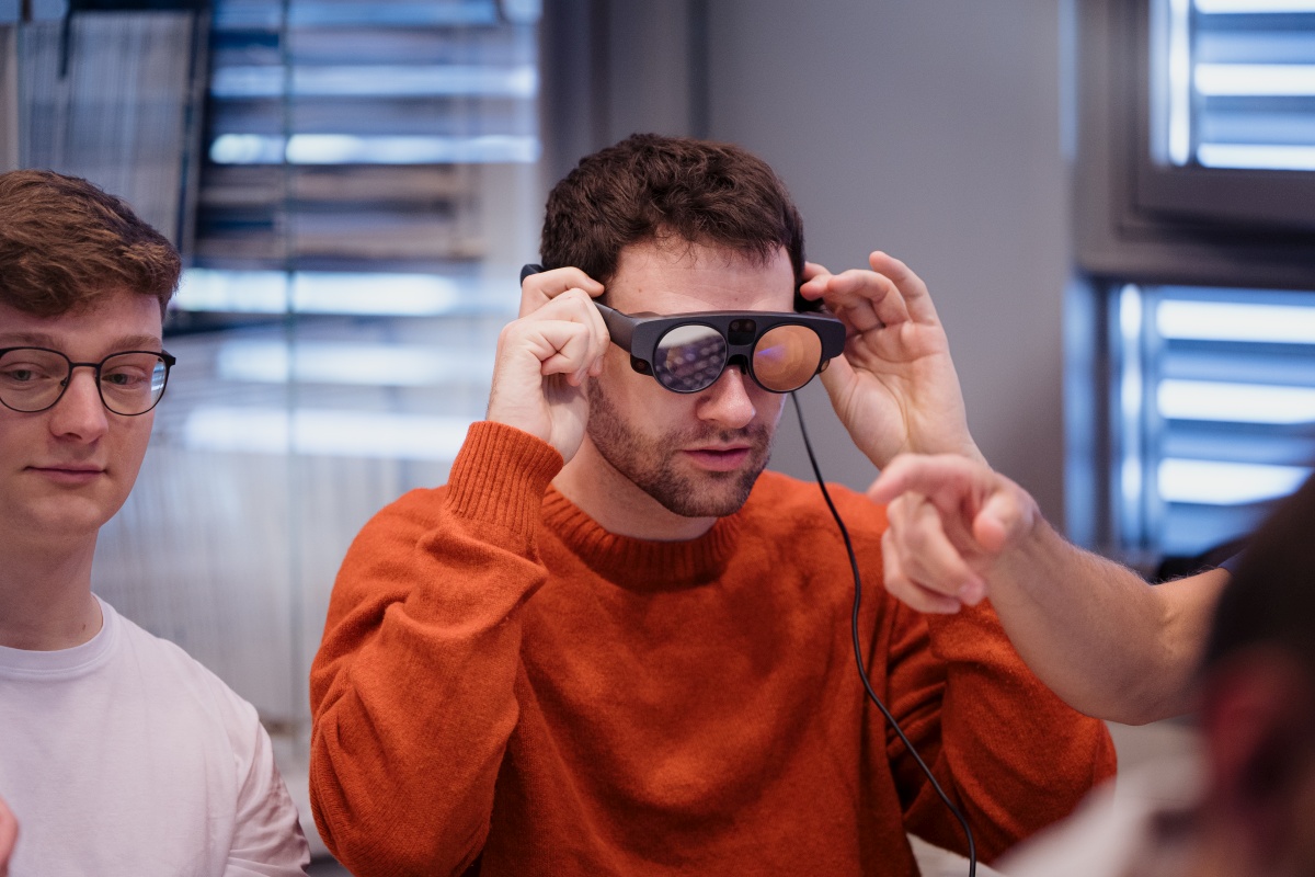
The students are already familiar with the technology from a course module taught by Professor Francesca Spadea, head of the Institute of Biomedical Engineering at KIT, who is also present in the operating room that day. In that module, the students had to program their own navigation system. Using MRI and CT data, they determined the position of a tumor – a simplified version of the system now being used with high precision in the operating room.
Precision Work on a Pulsating Brain
After lengthy preparation, everything happens very quickly: the first incision is made, the skin is opened, three small holes are drilled, and a rounded section of the skull is sawed out. Spetzger positions the surgical microscope over the patient’s head, and after opening the dura mater, the students see the pulsating brain on the screen. No one faints – and Spetzger carefully begins to remove the tumor tissue. Again and again, he places a small ultrasound probe directly on the brain. A new image appears on the screen in real time, showing how far the procedure has progressed. This is because the brain changes during surgery, shifting slightly – and with it, the orientation.
During the surgery, Brombach explains step by step what is happening. A student asks,
“How can you tell during removal which tissue is tumorous and which is healthy?” Brombach replies, “Tumor tissue often differs in color, consistency, and blood supply, but the boundaries are rarely well defined. That’s why, in addition to the surgeon’s experience, we also need the navigation system and ultrasound.”
A tissue sample is sent to the lab during the surgery. Later, the suspicion that the tumor is a glioblastoma is confirmed. The malignant tissue grows diffusely throughout the brain and cannot be completely removed. “After the surgery, the tumor will also need to be treated with radiation. I think this may allow us to give the patient several additional years of life,” says Brombach.
Stress That Pays Off
After about two hours, the tumor is removed: “Do you see that? The tumor was bigger than my thumb,” Spetzger says, gesturing for emphasis. As he takes off his surgical gown, he answers many more questions. The students thank him for the extraordinary opportunity before leaving the operating room and returning to their daily routine.
When asked about the student’s question from the lecture as to whether his job as a surgeon and lecturer is stressful, Spetzger replies with a laugh: “For me, it’s positive stress. I know former students who sat in my lecture six years ago and now work at major medical technology companies. That’s the best feedback.”
Leonie Kroll, August 6, 2026
Medicine of the Future
Issue 2/2026 of the research magazine lookKIT is dedicated to the theme of the Science Year 2026.
To the magazine