
New Imaging Technology Makes Corneal Nerves Visible Over a Large Area
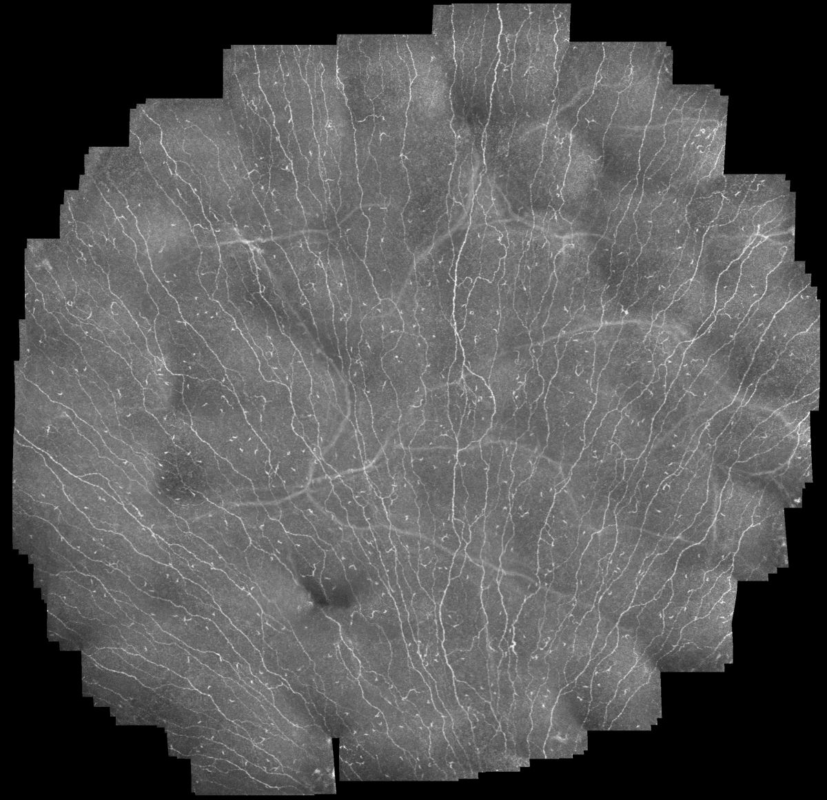
Diseases of the nerves can often only be detected once damage has already occurred. Researchers at the Karlsruhe Institute of Technology (KIT) and international partners have developed a new imaging platform that visualizes the nerve plexus of the eye’s cornea over a large area and at cellular resolution. The method increases the imaged area by a factor of 60 compared with previous methods and opens up new possibilities for the noninvasive examination of ocular diseases.
Corneal Nerves as a Panoramic Image
Unlike conventional microscopy, which captures only small areas of the cornea, the new method produces a “panoramic image” of the cornea that is assembled like a mosaic. “The combination of automated eye tracking, intelligent image acquisition, and computer-assisted image analysis has opened new possibilities for large-scale cellular imaging“, says Dr. Stephan Allgeier of the Institute for Automation and Applied Informatics (IAI) at KIT.
At the Institute for Automation and Applied Informatics (IAI), researchers developedthe automated gaze-guiding system during imaging and the algorithms for generating the panoramic images. The project partners are now transferring the system, jointly developed in Karlsruhe and Rostock, to Singapore for clinical studies.
The precise visualization of corneal nerves and the resident immune cells enables improved investigation of numerous neurodegenerative diseases and inflammatory diseases of the ocular surface, such as diabetic neuropathy. “Our vision is to detect nerve damage earlier using this noninvasive method so that therapeutic measures can be initiated at an early stage,” says Allgeier.
The project was funded by the German Research Foundation (DFG). Alongside KIT, the Singapore National Eye Centre, and the University Medical Center Rostock are participating.
lkr, August 13, 2026