
Every cell is enclosed by a thin double layer of lipids that separates the distinct internal environment of the cell from the extracellular space. Damage to this lipid bilayer, also referred to as plasma membrane, disturbs the cellular functions and may lead to the death of the cell. For example, downhill walking tears many little holes into the plasma membranes of the muscle cells in our legs. To prevent irreparable damage, muscle cells have efficient systems to seal these holes again. Researchers at Karlsruhe Institute of Technology (KIT) and Heidelberg University have succeeded for the first time in observing membrane repair in real-time in a living organism.
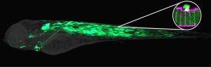
The results were published in the recent issue of the journal Developmental Cell. Using a novel high-resolution imaging method, Prof. Uwe Strähle and Dr. Urmas Roostalu for the first time observed membrane repair in real time in a living animal. They tagged repair proteins with fluorescent proteins in muscle of the transparent zebrafish larvae. With a laser, the researchers burned tiny holes into the plasma membrane of muscle cells and followed the repair of the holes under the microscope. They showed that membrane vesicles together with two proteins Dysferlin and Annexin A6 rapidly form a repair patch. Other Annexins accumulate subsequently on the injured membrane. These studies by Karlsruhe and Heidelberg researchers suggest that the cell assembles a multilayered repair patch from the inside that seals off the cell’s interior from the extracellular environment. Moreover, it was found that there is a specialized membrane area that supplies rapidly the membrane that is needed for sealing the plasma membrane hole.
This animal model for membrane repair will contribute to the identification of new proteins in this sealing process and will help elucidate the underlying mechanisms. The results may contribute to the development of therapies for human myopathies and open up new possibilities in biotechnology.
Literature:
Urmas Roostalu, Uwe Strähle: In Vivo Imaging of Molecular Interactions at Damaged Sarcolemma; Developmental Cell, 13 March 2012, Volume 22, Issue 3. http://www.cell.com/developmental-cell/current
In close partnership with society, KIT develops solutions for urgent challenges – from climate change, energy transition and sustainable use of natural resources to artificial intelligence, sovereignty and an aging population. As The University in the Helmholtz Association, KIT unites scientific excellence from insight to application-driven research under one roof – and is thus in a unique position to drive this transformation. As a University of Excellence, KIT offers its more than 10,000 employees and 22,800 students outstanding opportunities to shape a sustainable and resilient future. KIT – Science for Impact.