
How Fungi Grow: A Movie from Inside the Cell
Fungi forming mold on food are hazardous. Fungi supplying antibiotics are beneficial. Fungi may be harmful pathogens. On the other hand, they are used for the production of food or medicine and in bioengineering. In either case, it is required to precisely understand their growth mechanism. Researchers of Karlsruhe Institute of Technology (KIT) have made a big step forwards: Using high-performance light microscopy, they watched mold fungi as they grew in the cell. The findings are presented in Science Advances.
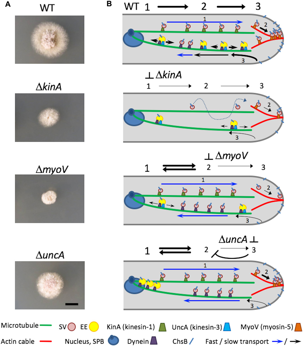
Like most fungi, mold fungi are hyphal fungi. They consist of filamentous cells, hyphae, which may form large networks, mycelia. The hyphae of about 3 µm in thickness exclusively grow by directed extension of their tips. They grow very rapidly, by about 1.5 mm per day. An important objective of biological fundamental research is to understand this growth on the molecular level, as hyphal growth plays an important role in both health-damaging effects and beneficial applications of fungi.
Movies show material transport in the fungal cell
For their studies, the researchers tagged a key enzyme required for building the chitin-containing cell wall with a fluorescent protein and observed the latter in the living cell with the help of high-resolution microscopy (nanoscopy). Use of ultrasensitive cameras in the microscope enabled high-speed imaging of tip growth and of the transport of individual vesicles. These images resemble small movies and allow to precisely determine transport speed of the vesicles. They reveal how building materials are packed into smallest vesicles and transported along the fiber structures of the cell skeleton to the cell tip by transport vehicles, the motor proteins. Motor proteins are very small nanomotors that dock to the fiber structures with two small “feet” and walk on these structures. Using genetically modified fungi, the scientists also identified the motor proteins responsible for the transports.
Findings contribute to combating pathogenic fungi
From their observations, the researchers of the Institute of Applied Physics and the Institute for Applied Biosciences of KIT derived a first comprehensive model to describe how the rapidly growing hyphal tip is supplied with construction material. This is an important step towards complete molecular understanding of directed cell growth processes, Professor Gerd Ulrich Nienhaus of KIT’s Institute of Applied Physics says. “The findings made in hyphal fungi are of general relevance to biology, as they can be transferred to other cells and organisms. On the other hand, they open up new opportunities to specifically influence fungal growth, which is important to the mitigation of pathogenic species in medicine.”
More info:
advances.sciencemag.org/content/4/1/e1701798
Go directly to the videos:
advances.sciencemag.org/content/suppl/2018/01/22/4.1.e1701798.DC1
rl, 05.02.2018