
Using a new X-ray technology, a research group has obtained hints as to how snakes lost their limbs in the course of evolution. With this, the scientists hope to further a debate among paleontologists: Do today’s snakes descend from ancient reptiles that lived on land or from marine animals? The detailed, three-dimensional X-ray images of the remains of a leg of an extinct snake reveal an inner architecture that is similar to that of today’s lizards. The results will be published in the Journal of Vertebrate Paleontology dated February 08.
The research project was directed by Alexandra Houssaye from the Muséum National d'Histoire Naturelle (MNHN), Paris (France). In addition, scientists from the European Synchrotron Radiation Facility (ESRF), Grenoble (France), where the X-ray images were taken, and from Karlsruhe Institute of Technology (KIT), where the recording technique and the corresponding instrument were developed, are involved.
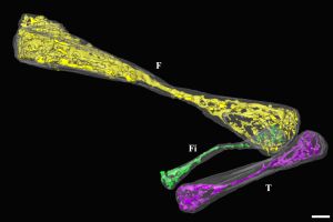
Only three snake fossils with preserved leg bones are known worldwide. During this experiment, an Eupodophis descouensi was studied. Its 95-million-year-old petrified remains were discovered in Lebanon ten years ago. The about 50 cm long snake has a small, 2 cm long leg in the pelvis area. This detail makes the fossil a key to understanding snake evolution, as it represents an intermediate phase, during which these snakes had not yet completely lost their legs inherited from the lizards. As only one leg is visible at the surface of the fossil, a second, hidden leg was expected to be hidden within the stone plate. It was detected by synchrotron radiation and imaged in all detail.
The highly resolved, three-dimensional images, particularly those of the leg hidden in the stone, suggest that this species had legs that grew less quickly or for a short time only. It is also evident from the images that the leg hidden in the stone was bent at the knee and possessed four ankle bones, but neither foot nor toe bones.
"Discovery of the inner structure of the hind limbs of Eupodophis now allows to investigate the process of regression of the limbs in snake evolution,” comments Alexandra Houssaye.
For the experiment, the researchers used synchrotron laminography. “This new technique was developed in particular for the investigation of larger, flat objects,” explains Lukas Helfen, who is delegated from the Institute for Synchrotron Radiation of Karlsruhe Institute of Technology to ESRF in order to equip and operate a beamline with this technology. “Synchrotron laminography resembles computer tomography (CT) that is used at many hospitals, but allows for a resolution in the micrometer range. This is a thousand times better than the resolution of a classical CT device.” During laminography, the fossil is rotated at an oblique angle to an intensive, high-energy X-ray beam, with thousands of two-dimensional images being recorded during the rotation from 0° to 360°. From these individual images, a highly resolved, three-dimensional model is calculated, which also discloses hidden details, such as the inner structure of the legs.
“Large synchrotrons can unveil microscopic details in fossils that remain hidden to other techniques. Moreover, synchrotrons can study these valuable objects in a non-destructive manner,” adds Paul Tafforeau from ESRF, one of the co-authors of the study.
Publication: A. Houssaye, F. Xu, L. Helfen, V. De Buffrénil, T. Baumbach, P. Tafforeau: Three-dimensional pelvis and limb anatomy of the Cenomanian hind-limbed snake Eupodophis descouensi (Squamata, Ophidia) revealed by synchrotron-radiation computed laminography. Journal of Vertebrate Paleontology 2011 31(1):1-6.
Contact data of the scientists involved:
Alexandra Houssaye
Departement Histoire de la Terre, Muséum National d'Histoire Naturelle
c/o Estelle Merceron
merceron ∂does-not-exist.mnhn fr
Paul Tafforeau
European Synchrotron Radiation Facility
BP 220, 6 rue Jules Horowitz
38043 Grenoble Cedex, France
paul tafforeau ∂does-not-exist.esrf fr
or
Claus Habfast
GSM +33 666 662 384
claus habfast ∂does-not-exist.esrf fr
Lukas Helfen
Institute for Synchrotron Radiation
Ångström Source Karlsruhe (ANKA)
Karlsruhe Institute of Technology
76021 Karlsruhe, Germany
lukas helfen ∂does-not-exist.kit edu
Being “The Research University in the Helmholtz Association”, KIT creates and imparts knowledge for the society and the environment. It is the objective to make significant contributions to the global challenges in the fields of energy, mobility, and information. For this, about 10,000 employees cooperate in a broad range of disciplines in natural sciences, engineering sciences, economics, and the humanities and social sciences. KIT prepares its 22,800 students for responsible tasks in society, industry, and science by offering research-based study programs. Innovation efforts at KIT build a bridge between important scientific findings and their application for the benefit of society, economic prosperity, and the preservation of our natural basis of life. KIT is one of the German universities of excellence.