
A group of KIT scientists headed by Professor Gerd Ulrich Nienhaus has developed a new fluorescent marker protein. The fluorescent light of the photoactivatable protein “mIrisFP” can be switched on and off and its color of emission can be changed from green to red. The protein allows for dynamic studies of cells and organisms and opens up new opportunities for cell biology and molecular medicine research. This development has just been presented in the journal “Nature Methods”.
Fluorescent proteins re-emit incident light efficiently, so that they are visible in living cells. As genetically encoded fluorescent markers, they can be used in a diverse range of experiments in the life sciences. If the gene of a certain protein is extended by the genetic information (DNA sequence) for a fluorescent protein, the cell produces a so-called fusion protein that can be detected due to its characteristic fluorescence.
The green fluorescent protein (GFP) of a pacific jellyfish was the first and, for a long time, the only one known. In recent years, researchers have discovered fluorescent proteins in other invertebrate marine fauna and also identified photactivatable proteins, the fluorescence of which can be controlled specifically by light irradiation. Natural forms of these proteins, however, are not suited for biotechnology applications; they first have to be optimized by often time-consuming protein engineering. So far, the fluorescence of such marker proteins could either be switched on and off, or the color of the light could be changed.
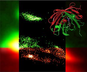
The group of KIT researchers headed by Gerd Ulrich Nienhaus from the DFG Center for Functional Nanostructures (CFN) has now introduced a marker protein called “mIrisFP” in cooperation with scientists from the University of Ulm, the University of Southampton/UK, and the University of Illinois/USA. This protein combines two activation modes that can be controlled by light, irreversible photoconversion from green to red and reversible on/off switching of the fluorescence. In addition, mIrisFP is monomeric, i.e., it consists of a single unit only; it does not form molecular complexes.
“As a result, mIrisFP is excellently suited for life science applications,” explains Professor Nienhaus. “Dynamic investigations in living cells and organisms are particularly exciting.” By specific light irradiation, certain cells in early embryonal stages of model organisms can be marked and tracked over days and weeks. Specific marking of cell compartments is achieved with signal peptides that are attached to the fluorescent protein by genetic modification on the DNA level. Fusion constructs of a fluorescent marker protein with a protein under investigation are of particular importance for making the protein visible. Photoactivatable fluorescent proteins also play a key role in highest-resolution fluorescence microscopy.
With mIrisFP, cells can be imaged with spatial resolutions of 20 to 30 nm, which is far below Abbe’s resolution limit of about 200 nm that was considered the physical limit for a long time and prevented detailed insights into molecular processes. In the journal “Nature Methods”, the KIT researchers present the new marker protein and study molecular processes during the movement of a human cancer cell by means of highest-resolution photoactivation localization microscopy (PALM). In this way, they were able to observe that certain proteins visualized with mIrisFP are removed from one area of the cell and are subsequently integrated into new structures in another area of the cell.
Literature
A photoactivatable marker protein for pulse-chase imaging with superresolution. Jochen Fuchs, Susan Boehme, Franz Oswald, Per Niklas Hedde, Maike Krause, Jörg Wiedenmann & G Ulrich Nienhaus. Nature Methods. Published online: 4 July 2010; | doi: 10.1038/nmeth.1477.
http://dx.doi.org/_10.1038/nmeth.1477
Being “The Research University in the Helmholtz Association”, KIT creates and imparts knowledge for the society and the environment. It is the objective to make significant contributions to the global challenges in the fields of energy, mobility, and information. For this, about 10,000 employees cooperate in a broad range of disciplines in natural sciences, engineering sciences, economics, and the humanities and social sciences. KIT prepares its 22,800 students for responsible tasks in society, industry, and science by offering research-based study programs. Innovation efforts at KIT build a bridge between important scientific findings and their application for the benefit of society, economic prosperity, and the preservation of our natural basis of life. KIT is one of the German universities of excellence.